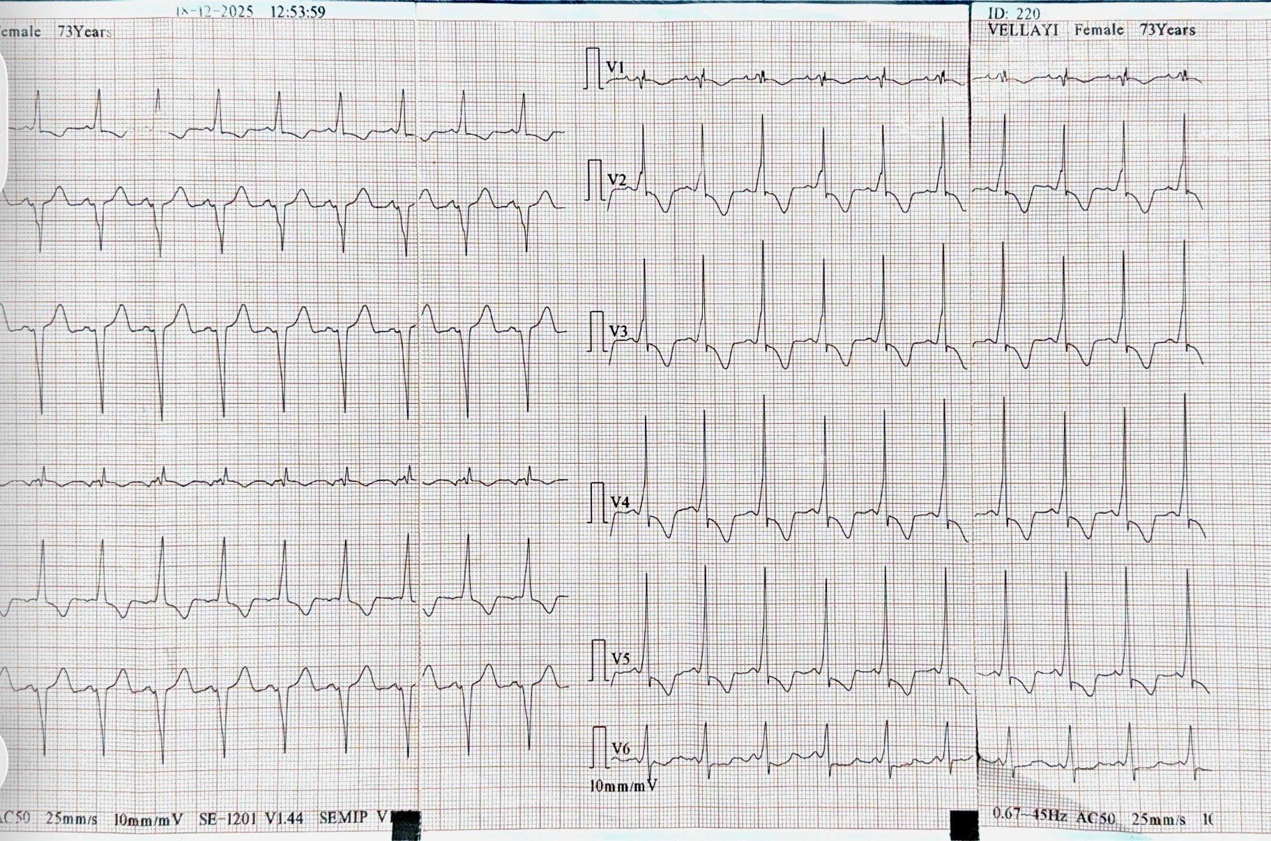
The ECG demonstrates sinus tachycardia, with subtle P waves merging into the preceding T waves (as shown in the image).
There is right axis deviation, QRS prolongation (>100 ms), and a distinct terminal R wave in aVR (also highlighted in the image).
The QT interval is prolonged as well.
Although ST elevation in aVR and V1 may raise concern for acute MI, and a wide QRS can occur in hyperkalemia, the overall ECG pattern, combined with the patient’s symptoms (decreased level of consciousness and severely low blood pressure) strongly suggests sodium channel blocker toxicity, most classically TCA overdose given the patient’s history of depression , or toxicity from certain class I antiarrhythmic agents.